• Home
  • Dr. Lewis
    • Curriculum Vitae
  • Staff
  • Premium Cataract Surgery
  • Locations
    • Surgery Center
  • Testimonials
  • Contact
  • Blog
  • Patient Form

CataractPhiladelphia

Cataract Surgery Philadelphia

  • Cataracts
  • Complex
    • Cataracts and Astigmatism
    • Cataracts and Glaucoma
      • Hydrus MicroStent: Cataracts and Glaucoma
      • iStent
      • AquaFlow
      • Ahmed M4
    • Cataracts and Cornea
    • Cataracts and Dry Eye
    • Cataracts after LASIK
    • After Retinal Surgery
    • Cataracts After Glaucoma Surgery
    • Cataracts in Monocular Patients
    • Cataracts after Trauma
    • Cataracts in Special Populations
    • Cataract Instrumentation
    • Cataract Failures
  • Implants
    • Premium Implants
    • RxSight Light Adjustable Lens
    • PanOptix Trifocal
    • AcrySof IQ Vivity
    • Toric Implants
  • Co-Management
  • Risks
  • Yag

Macular Edema Following Intravitreal Triamcinolone as an Alternative to Post Cataract Anti-inflammatory Drops

James S. Lewis, MD
Elkins Park, PA

ABSTRACT

To investigate the incidence of macular edema following uncomplicated cataract surgery in which intravitreal triamcinolone is injected in lieu of post-operative topical steroids and NSAIDs.

BACKGROUND

Dropless cataract surgery has been proposed as an alternative to the standard of care. Presently, patients self-administer topical antibiotics, steroids and NSAIDs multiple times a day for one to eight weeks. The impetus for this investigation includes rampant medication non-compliance, excessive branded drug costs, non-physician initiated pharmaceutical substitution, increased generic drug prices, uncertain bioavailability, excess toxicity, erratic formulary coverage policies, and the deliberate and drastic reduction in branded pharmaceutical sampling.

Ophthalmologists, optometrists and office staff have become a virtual sales force for the pharmaceutical industry navigating the drug procurement process. This includes negotiating the compromise between the high cost of drops and patient financial resources, amending the electronic medical record when the doctor’s first choice is rejected, and responding to unpredictable coverage adjudication and other inefficiencies which disrupt our present system.

In an era in which public health optimization vies with individual patient care and physicians are mandated to work more efficiently with fewer resources, responsible health care providers must find an alternative to this untenable system. A cataract procedure without post-operative drops has been the surgeon’s goal for decades.

Macular Edema after Dropless Cataract Surgery

METHODS AND MATERIALS

200 consecutive cataract procedures of 129 patients without pre-existing macular pathology from a single surgeon private practice were imaged with retinal OCT pre-operatively and at one day, one week, one month, and two months following uncomplicated surgery. Following intraocular lens implantation and prior to visocelastic removal a transzonular instillation of 0.2cc of a commercially available compounded medication* containing triamcinolone acetonide was performed. The central foveal thickness (CFT) was recorded in all cases at the intervals described above.

RESULTS

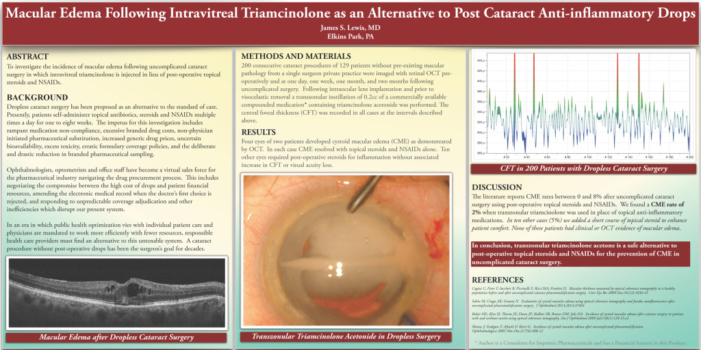
Four eyes of two patients developed cystoid macular edema (CME) as demonstrated by OCT. In each case CME resolved with topical steroids and NSAIDs alone. Ten other eyes required post-operative steroids for inflammation without associated increase in CFT or visual acuity loss.

Transzonular Triamcinolone Acetonide in Dropless Surgery
CFT in 200 Patients with Dropless Cataract Surgery

DISCUSSION

The literature reports CME rates between 0 and 8% after uncomplicated cataract surgery using post-operative topical steroids and NSAIDs. We found a CME rate of 2% when transzonular triamcinolone was used in place of topical anti-inflammatory medications. In ten other cases (5%) we added a short course of topical steroid to enhance patient comfort. None of these patients had clinical or OCT evidence of macular edema.

CONCLUSION

In conclusion, transzonular triamcinolone acetone is a safe alternative to post-operative topical steroids and NSAIDs for the prevention of CME in uncomplicated cataract surgery.

REFERENCES

Cagini C; Fiore T; Iaccheri B; Piccinelli F; Ricci MA; Fruttini D. Macular thickness measured by optical coherence tomography in a healthy population before and after uncomplicated cataract phacoemulsification surgery. Curr Eye Res 2009 Dec;34(12):1036-41  

Sahin M; Cingu AK; Gozum N. Evaluation of cystoid macular edema using optical coherence tomography and fundus autofluorescence after uncomplicated phacoemulsification surgery. J Ophthalmol 2013;2013:37601

Belair ML; Kim SJ; Thorne JE; Dunn JP; Kedhar SR; Brown DM; Jabs DA. Incidence of cystoid macular edema after cataract surgery in patients with and without uveitis using optical coherence tomography. Am J Ophthalmol 2009 Jul;148(1):128-35.e2

Mentes J; Erakgun T; Afrashi F; Kerci G. Incidence of cystoid macular edema after uncomplicated phacoemulsification. Ophthalmologica 2003 Nov-Dec;217(6):408-12

* Author is a Consultant for Imprimis Pharmaceuticals and has a Financial Interest in this Product.

CALL FOR AN APPOINTMENT

(215) 886-9090

RxSight Light Adjustable Lens

Select uncompromised clarity with an IOL that matches your visual requirements

learn more

PanOptix Trifocal dominates Cataract Philadelphia

Enjoy a glasses-free life, with the most advanced multifocal IOL.

learn more

AcrySof IQ Vivity

Designed to deliver an extended focal range and a monofocal-like visual disturbance profile

learn more

Dropless and Less Drops Cataract Surgery

Choose safer surgery and save $100 to $300 per eye

learn more

Hydrus MicroStent: Cataracts and Glaucoma

Stop your glaucoma drops with the Hydrus MicroStent.

learn more

Live Surgery Stream

lewislasik

Full compliance with all guidelines 😷 related to the Coronavirus crisis

Lewis LASIK
Our practice will start seeing patients again on M Our practice will start seeing patients again on Monday, May 11th in full compliance with all local, state, and federal guidelines. This includes masks, social distancing, and the disinfection of all common surfaces. Elective surgeries are also being rescheduled. Care will be taken to avoid office congestion and minimize exposure to COVID-19.

We require: ➡️ You do not have fever, chills, shaking, muscle pain, headache, sore throat, or a loss of taste or smell ➡️ You have not had exposure to anyone with a flu-like illness within the past two weeks ➡️ You wear a mask or equivalent facial covering over both your nose and mouth ➡️ You agree to maintain social distancing ➡️ You avoid touching your eyes, nose, mouth, and face ➡️ You enter the office alone if possible

Expect to hear from our staff shortly. You may contact us at your convenience from links at jameslewismd.com
Chorioretinal folds are a known finding following Chorioretinal folds are a known finding following penetrating glaucoma surgery, as in these two cases who underwent Ahmed valve tube shunt placement. Prevalence is estimated between 10-50% of incisional glaucoma surgeries.

Pic 1.) several linear chorioretinal folds throughout the posterior pole. Intraocular pressure was 4mmHg at the time of this photo. The fundus and visual acuity returned to baseline within a week as IOP leveled at 10mmHg.
Pic 2.) Small choroidal folds can be seen distributed temporal to the macula. This image also demonstrates a large hemorrhage consistent with ocular decompression retinopathy.
Pic 3.) shows complete resolution of the choroidal folds and hemorrhage after 4 weeks in patient 2.

#ophthalmology #ophthalmologist #ophthalmictech #ophthalmologyresident #ophthalmicphotography #ophthalmicsurgery #cornea #corneasurgery #eyesurgeon #eyesurgery #eye #oculardisease #optometry  #optometrystudent #optom #sunyoptometry #osuopt #salusuniversity #glaucoma #glaucomasurgery #retina #chorioretinalfolds
Inflammation of the anterior chamber can create fi Inflammation of the anterior chamber can create fibrin plaques that are readily seen within the pupil. The second and third images demonstrate an almost completely occluded pupil with synechia formation. The fourth image demonstrates an ultrasound biomicroscopy image of a patient in angle closure following complete pupil occlusion from fibrin (blue arrow). Aggressive corticosteroid therapy can ‘melt’ the fibrin and cycloplegics can mechanically disrupt it. Nd:YAG laser can also instantly disrupt total occlusion.

#ophthalmology #ophthalmologist #ophthalmictech #ophthalmologyresident #ophthalmicphotography #ophthalmicsurgery #cornea #corneasurgery #eyesurgeon #eyesurgery #eye #oculardisease #optometry #ocularinflammation #optometrystudent #optom #sunyoptometry #osuopt #salusuniversity
Descemet stripping endothelial keratoplasty (DSEK) Descemet stripping endothelial keratoplasty (DSEK) is a corneal transplant procedure that replaces only the innermost cells of the cornea. It is readily combined with cataract surgery to improve refractive outcomes. This is a one day post operative visit of a DSEK showing faint edema and remaining air bubble. The air bubble will typically dissolve over the first 48-72 hours.

#ophthalmology #ophthalmologist #ophthalmictech #ophthalmologyresident #ophthalmicphotography #ophthalmicsurgery #cornea #corneasurgery #eyesurgeon #eyesurgery #corneatransplant #dsek #fuchsdystrophy #endothelium #eye #oculardisease #optometry #optometrystudent #optom #sunyoptometry #osuopt #salusuniversity
Ocular surface disease is a complex state that rep Ocular surface disease is a complex state that represents a poorly performing pre-corneal tear film. It can frustrate many patients and can be challenging for clinicians. No two cases are alike.

In this case, a young patient without evidence of desiccation or inflammation had persistent physical symptoms. Biomicroscopy revealed clear lid margins, no conjunctival injection, and no corneal epitheliopathy. Meibography (imaging the meibomian glands) provides insight into the cause. A previous course of oral medication that acts on sebaceous glands has likely impacted his meibomian glands as well. The gland dropout is easily seen as truncation and atrophy. The final video demonstrates a noninvasive tear-breakup time where the red squares are regions with early loss of tear film stability.

#ophthalmology #ophthalmologist #ophthalmictech #ophthalmologyresident #ophthalmicphotography #ophthalmicsurgery #cornea #corneasurgery #eyesurgeon #eyesurgery #dryeye #ocularsurfacedisease #OSD #eye #oculardisease #optometry #optometrystudent #optom #sunyoptometry #osuopt #salusuniversity
Iris cysts are benign. If suspected, they should b Iris cysts are benign. If suspected, they should be imaged with ultrasound biomicroscopy (UBM) to confirm the diagnosis and rule out other causes of iris elevation.
#ophthalmology #ophthalmologist #ophthalmictech #ophthalmologyresident #ophthalmicphotography #ophthalmicsurgery #cornea #corneasurgery #eyesurgeon #eyesurgery #ubm #eye #oculardisease #optometry #optometrystudent #optom #sunyoptometry #osuopt #salusuniversity
There are various types of cornea transplantation There are various types of cornea transplantation procedures and each is best suited for different patients based on diagnosis and other factors. This is an MDALK (maximum depth anterior lamellar keratoplasty) that retains the innermost layer of cells and membrane, but replaces as much diseased tissue as possible. It has excellent clarity and comparatively good visual outcomes. 
#ophthalmology #ophthalmologist #ophthalmictech #ophthalmologyresident #ophthalmicphotography #ophthalmicsurgery #cornea #corneasurgery #eyesurgeon #eyesurgery #eye #oculardisease #optometry #optometrystudent #optom #sunyoptometry #osuopt #salusuniversity #mdalk #corneatransplant
Corneal infection! This series of images highlight Corneal infection! This series of images highlights a case of pseudomonas aeruginosa microbial keratitis (corneal infection) related to contact lens wear. Keep in mind the cornea is about 0.5 millimeters thick! Not much room to allow an infection to move deeper.
Pic 1. The resultant scar after the infection cleared.
Pic 2. At presentation, the eye is injected (red) with a large central infiltrate (immune response) and necrotic stromal tissue (dying cells). Pic 3. There was extensive inflammation in the anterior chamber including fibrin strands
Pic 4. Note the presence of a hypopyon (accumulation of white blood cells at the bottom of the anterior chamber).
Pic 5. This is two days after initiating treatment with fortified antibiotics. The pupil is intentionally dilated (for comfort) and anterior chamber inflammation is improving. There is a small unrelated fiber present that was subsequently removed.
Pic 6. A closer look at the scar and the shadow it casts. These scars disrupt the vision permanently. Fortunately this patient has achieved good acuity, yet blur, glare and haze can be persistent.
(Sorry for poor photo quality in pictures 2/3/4)

It is important to work quickly to identify the organism responsible for these infections to tailor treatment appropriately. If left untreated infection can spread to devastating results.

#ophthalmology #ophthalmologist #ophthalmictech #ophthalmologyresident #ophthalmicphotography #ophthalmicsurgery #cornea #corneasurgery #eyesurgeon #eyesurgery #microbialkeratitis #eye #oculardisease #optometry #optometrystudent #optom #sunyoptometry #osuopt #salusuniversity
Load More... Follow on Instagram

At the Philadelphia, Pennsylvania cataract surgery offices of Dr. James S. Lewis, patients are assured the highest quality treatments and impeccable care. Dr. Lewis and his team use the latest surgical technologies and techniques to deliver the best possible results. CataractPhiladelphia offers a wide range of premium IOLs, including PanOptix, Vivity, Toric and Light Adjustable Lens. Dr. Lewis also treats patients with both cataracts and glaucoma through an enhanced range of micro-invasive glaucoma surgical options.

loader-image
Salus University
ISRS
ASCRS
American Academy of Ophthalmology
ESCRS
AECOS

* currently only PanOptix, Vivity, Toric, and the Light Adjustable Lens patients qualify for ONCE AND DONE.

 
RELATED LINKS
  • Fab Eye Care Center
  • Lewis LASIK
  • Dry Eye Philadelphia
  • Laser Dry Eye
  • LASIK TV

CALL FOR AN APPOINTMENT

(215) 886-9090

8380 Old York Road
Suite 110
Elkins Park, PA 19027

Copyright © 2021 CataractPhiladelphia.com | website: digitalpaintbrush.com Log in