• Home
  • Dr. Lewis
    • Curriculum Vitae
  • Staff
  • Premium Cataract Surgery
  • Locations
    • Surgery Center
  • Testimonials
  • Contact
  • Blog
  • Patient Form

CataractPhiladelphia

Cataract Surgery Philadelphia

  • Cataracts
  • Complex
    • Cataracts and Astigmatism
    • Cataracts and Glaucoma
      • Hydrus MicroStent: Cataracts and Glaucoma
      • iStent
      • AquaFlow
      • Ahmed M4
    • Cataracts and Cornea
    • Cataracts and Dry Eye
    • Cataracts after LASIK
    • After Retinal Surgery
    • Cataracts After Glaucoma Surgery
    • Cataracts in Monocular Patients
    • Cataracts after Trauma
    • Cataracts in Special Populations
    • Cataract Instrumentation
    • Cataract Failures
  • Implants
    • Premium Implants
    • RxSight Light Adjustable Lens
    • PanOptix Trifocal
    • AcrySof IQ Vivity
    • Toric Implants
  • Co-Management
  • Risks
  • Yag

AquaFlow: Cataracts and Glaucoma – James S. Lewis, MD

Serving Northeast Philadelphia, King of Prussia, Montgomery County, Conshohocken, Philadelphia, Phoenixville and the Main Line


AquaFlow™ or more generally, non-penetrating deep sclerectomy (NPDS) combined with cataract surgery is an excellent option for patients with cataracts and glaucoma.

Some Philadelphia / Bucks County cataract patients are content to continue using their glaucoma medications following cataract surgery. Unfortunately, the cost of drops and co-payments continue to rise. Furthermore glaucoma medications have systemic side effects and after years patients can develop tachyphylaxis (less effective over time) and allergy to these medications. Also, immediate post-operative pressure spikes in cataract patients with glaucoma can have significant unwanted consequences. These factor motivate skilled cataract surgeons to combine cataract surgery with minimally invasive glaucoma surgery (MIGS). Similarly, many cataracts and glaucoma patients prefer their surgeon address both conditions at the time for surgery. In some cases a combined surgical procedure is absolutely necessary to maintain ocular health.

Non-penetrating deep sclerectomy (NPDS) combined with cataract surgery is an excellent choice for patients with cataracts and glaucoma. Alternative procedures like trabeculectomy, mitomycin trabeculectomy, Express Shunt, Ahmed Value, Molteno Tube and other setons significantly delay visual rehabilitation after cataract surgery. Dr. James Lewis has been performing combined cataract and NPDS for almost a decade. In particular, he uses the AquaFlow implant to maintain excellent glaucoma pressure control after cataract surgery. Visual recovering is almost immediate and comfort is maintained. Unlike full thickness glaucoma procedures, no excessive pressure fluctuations, flat chambers, choroidals, choroidals hemorrhages, macular edema or retinal hypotony occur. Cataract extraction and Aquaflow Surgery as a combined procedure takes less than 30 minutes, is performed under local anesthesia, uses no sutures, has no foreign body sensation and does not involve cosmetic abnormalities resulting from removal of iris tissue. (Almost all other glaucoma procedures involve removing small portions of the iris often distorting the size and shape of the pupil.)

Dr. Lewis has performed thousands of these procedures and is the most experienced Aquaflow Surgeon in the United States.

For patients with less aggressive glaucoma, Dr. Lewis may choose to combine cataract surgery with iStent.

Your browser does not support the video tag or the file format of this video. https://www.webestools.com/
  • Glaucoma Introduction
  • Glaucoma Description
  • How Fluid Circulates in the Eye
  • Glaucoma Cause
  • Glaucoma Risk
  • Ocular Hypertension
  • Open-Angle Glaucoma
  • Narrow- Angle Glaucoma

 
Nearly one out of six patients of cataract age have some form of glaucoma. Sometimes this has not been diagnosed because glaucoma is a silent condition.

The region marked A is the cataract. Excess pressure causing glaucoma is produced at B. Their proximity help explain why cataracts and glaucoma frequently co-exist. Schlemm’s canal is the exit route for excess aqueous humor C and is the focus of the Aquaflow procedure.
 

The AquaFlow Procedure

Localize Schlemm’s Canal

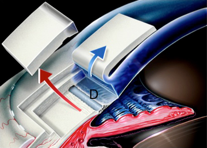
Immediately after the cataract is removed and the intraocular lens implanted Dr. Lewis exposes Schlemm’s canal (D) deep in the stroma. Notice the entire surgery takes place without actually entering the eye consequently the name, non-penetrating deep sclerectomy.

Unroofing Schlemm’s Canal

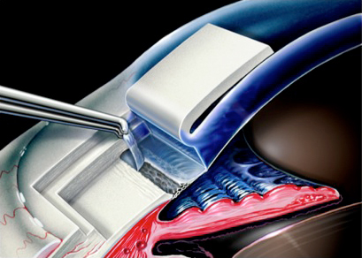
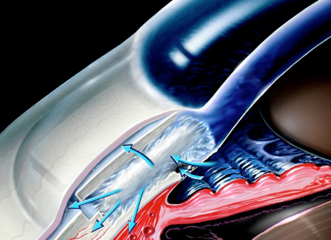
Excess aqueous humor fails to flow though Schleem’s canal in patients with glaucoma. For this reason the canal is opened allowing more fluid to pass from the anterior chamber of the eye, through the trabecular meshwork and back into the normal ocular channels.

Fig 5

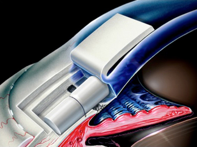
Inserting the AquaFlow Device
The AquaFlow shunt acts as a stent allowing fluid to flow into a tiny natural reservoir hidden below the ocular surface behind the lid. There are no scars or filtering blebs to call attention to this very effective surgery.

Fig 6

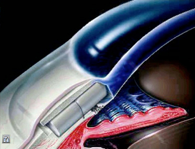
The AquaFlow Device Dissolves in 9 months
The AquaFlow is made of collagen and dissolves after it has created a new pathway for the excess intraocular pressure. Most of our Philadelphia cataract and glaucoma patients maintain excellent intraocular pressure control following AquaFlow insertion. Furthermore, many of them are able to achieve this without any glaucoma medications.

In addition to helping cataracts patients with glaucoma, Dr. Lewis is also experienced in treating cataract patients suffering from astigmatism and dry eye. Dr. Lewis is one of the more experienced cataracts surgeons in the United States, and he has been recognized as a foremost Philadelphia multifocal IOL (ReSTOR, Tecnis Multifocal and ReZoom), astigmatism-correcting IOLs (Crystalens Toric {Trulign} and Alcon Toric), and accommodating IOLs (Crystalens AO) provider.

Staar Surgical AquaFlow

CALL FOR AN APPOINTMENT

(215) 886-9090

RxSight Light Adjustable Lens

Select uncompromised clarity with an IOL that matches your visual requirements

learn more

PanOptix Trifocal dominates Cataract Philadelphia

Enjoy a glasses-free life, with the most advanced multifocal IOL.

learn more

AcrySof IQ Vivity

Designed to deliver an extended focal range and a monofocal-like visual disturbance profile

learn more

Dropless and Less Drops Cataract Surgery

Choose safer surgery and save $100 to $300 per eye

learn more

Hydrus MicroStent: Cataracts and Glaucoma

Stop your glaucoma drops with the Hydrus MicroStent.

learn more

Live Surgery Stream

lewislasik

Full compliance with all guidelines 😷 related to the Coronavirus crisis

Lewis LASIK
Our practice will start seeing patients again on M Our practice will start seeing patients again on Monday, May 11th in full compliance with all local, state, and federal guidelines. This includes masks, social distancing, and the disinfection of all common surfaces. Elective surgeries are also being rescheduled. Care will be taken to avoid office congestion and minimize exposure to COVID-19.

We require: ➡️ You do not have fever, chills, shaking, muscle pain, headache, sore throat, or a loss of taste or smell ➡️ You have not had exposure to anyone with a flu-like illness within the past two weeks ➡️ You wear a mask or equivalent facial covering over both your nose and mouth ➡️ You agree to maintain social distancing ➡️ You avoid touching your eyes, nose, mouth, and face ➡️ You enter the office alone if possible

Expect to hear from our staff shortly. You may contact us at your convenience from links at jameslewismd.com
Chorioretinal folds are a known finding following Chorioretinal folds are a known finding following penetrating glaucoma surgery, as in these two cases who underwent Ahmed valve tube shunt placement. Prevalence is estimated between 10-50% of incisional glaucoma surgeries.

Pic 1.) several linear chorioretinal folds throughout the posterior pole. Intraocular pressure was 4mmHg at the time of this photo. The fundus and visual acuity returned to baseline within a week as IOP leveled at 10mmHg.
Pic 2.) Small choroidal folds can be seen distributed temporal to the macula. This image also demonstrates a large hemorrhage consistent with ocular decompression retinopathy.
Pic 3.) shows complete resolution of the choroidal folds and hemorrhage after 4 weeks in patient 2.

#ophthalmology #ophthalmologist #ophthalmictech #ophthalmologyresident #ophthalmicphotography #ophthalmicsurgery #cornea #corneasurgery #eyesurgeon #eyesurgery #eye #oculardisease #optometry  #optometrystudent #optom #sunyoptometry #osuopt #salusuniversity #glaucoma #glaucomasurgery #retina #chorioretinalfolds
Inflammation of the anterior chamber can create fi Inflammation of the anterior chamber can create fibrin plaques that are readily seen within the pupil. The second and third images demonstrate an almost completely occluded pupil with synechia formation. The fourth image demonstrates an ultrasound biomicroscopy image of a patient in angle closure following complete pupil occlusion from fibrin (blue arrow). Aggressive corticosteroid therapy can ‘melt’ the fibrin and cycloplegics can mechanically disrupt it. Nd:YAG laser can also instantly disrupt total occlusion.

#ophthalmology #ophthalmologist #ophthalmictech #ophthalmologyresident #ophthalmicphotography #ophthalmicsurgery #cornea #corneasurgery #eyesurgeon #eyesurgery #eye #oculardisease #optometry #ocularinflammation #optometrystudent #optom #sunyoptometry #osuopt #salusuniversity
Descemet stripping endothelial keratoplasty (DSEK) Descemet stripping endothelial keratoplasty (DSEK) is a corneal transplant procedure that replaces only the innermost cells of the cornea. It is readily combined with cataract surgery to improve refractive outcomes. This is a one day post operative visit of a DSEK showing faint edema and remaining air bubble. The air bubble will typically dissolve over the first 48-72 hours.

#ophthalmology #ophthalmologist #ophthalmictech #ophthalmologyresident #ophthalmicphotography #ophthalmicsurgery #cornea #corneasurgery #eyesurgeon #eyesurgery #corneatransplant #dsek #fuchsdystrophy #endothelium #eye #oculardisease #optometry #optometrystudent #optom #sunyoptometry #osuopt #salusuniversity
Ocular surface disease is a complex state that rep Ocular surface disease is a complex state that represents a poorly performing pre-corneal tear film. It can frustrate many patients and can be challenging for clinicians. No two cases are alike.

In this case, a young patient without evidence of desiccation or inflammation had persistent physical symptoms. Biomicroscopy revealed clear lid margins, no conjunctival injection, and no corneal epitheliopathy. Meibography (imaging the meibomian glands) provides insight into the cause. A previous course of oral medication that acts on sebaceous glands has likely impacted his meibomian glands as well. The gland dropout is easily seen as truncation and atrophy. The final video demonstrates a noninvasive tear-breakup time where the red squares are regions with early loss of tear film stability.

#ophthalmology #ophthalmologist #ophthalmictech #ophthalmologyresident #ophthalmicphotography #ophthalmicsurgery #cornea #corneasurgery #eyesurgeon #eyesurgery #dryeye #ocularsurfacedisease #OSD #eye #oculardisease #optometry #optometrystudent #optom #sunyoptometry #osuopt #salusuniversity
Iris cysts are benign. If suspected, they should b Iris cysts are benign. If suspected, they should be imaged with ultrasound biomicroscopy (UBM) to confirm the diagnosis and rule out other causes of iris elevation.
#ophthalmology #ophthalmologist #ophthalmictech #ophthalmologyresident #ophthalmicphotography #ophthalmicsurgery #cornea #corneasurgery #eyesurgeon #eyesurgery #ubm #eye #oculardisease #optometry #optometrystudent #optom #sunyoptometry #osuopt #salusuniversity
There are various types of cornea transplantation There are various types of cornea transplantation procedures and each is best suited for different patients based on diagnosis and other factors. This is an MDALK (maximum depth anterior lamellar keratoplasty) that retains the innermost layer of cells and membrane, but replaces as much diseased tissue as possible. It has excellent clarity and comparatively good visual outcomes. 
#ophthalmology #ophthalmologist #ophthalmictech #ophthalmologyresident #ophthalmicphotography #ophthalmicsurgery #cornea #corneasurgery #eyesurgeon #eyesurgery #eye #oculardisease #optometry #optometrystudent #optom #sunyoptometry #osuopt #salusuniversity #mdalk #corneatransplant
Corneal infection! This series of images highlight Corneal infection! This series of images highlights a case of pseudomonas aeruginosa microbial keratitis (corneal infection) related to contact lens wear. Keep in mind the cornea is about 0.5 millimeters thick! Not much room to allow an infection to move deeper.
Pic 1. The resultant scar after the infection cleared.
Pic 2. At presentation, the eye is injected (red) with a large central infiltrate (immune response) and necrotic stromal tissue (dying cells). Pic 3. There was extensive inflammation in the anterior chamber including fibrin strands
Pic 4. Note the presence of a hypopyon (accumulation of white blood cells at the bottom of the anterior chamber).
Pic 5. This is two days after initiating treatment with fortified antibiotics. The pupil is intentionally dilated (for comfort) and anterior chamber inflammation is improving. There is a small unrelated fiber present that was subsequently removed.
Pic 6. A closer look at the scar and the shadow it casts. These scars disrupt the vision permanently. Fortunately this patient has achieved good acuity, yet blur, glare and haze can be persistent.
(Sorry for poor photo quality in pictures 2/3/4)

It is important to work quickly to identify the organism responsible for these infections to tailor treatment appropriately. If left untreated infection can spread to devastating results.

#ophthalmology #ophthalmologist #ophthalmictech #ophthalmologyresident #ophthalmicphotography #ophthalmicsurgery #cornea #corneasurgery #eyesurgeon #eyesurgery #microbialkeratitis #eye #oculardisease #optometry #optometrystudent #optom #sunyoptometry #osuopt #salusuniversity
Load More... Follow on Instagram

At the Philadelphia, Pennsylvania cataract surgery offices of Dr. James S. Lewis, patients are assured the highest quality treatments and impeccable care. Dr. Lewis and his team use the latest surgical technologies and techniques to deliver the best possible results. CataractPhiladelphia offers a wide range of premium IOLs, including PanOptix, Vivity, Toric and Light Adjustable Lens. Dr. Lewis also treats patients with both cataracts and glaucoma through an enhanced range of micro-invasive glaucoma surgical options.

loader-image
Salus University
ISRS
ASCRS
American Academy of Ophthalmology
ESCRS
AECOS

* currently only PanOptix, Vivity, Toric, and the Light Adjustable Lens patients qualify for ONCE AND DONE.

 
RELATED LINKS
  • Fab Eye Care Center
  • Lewis LASIK
  • Dry Eye Philadelphia
  • Laser Dry Eye
  • LASIK TV

CALL FOR AN APPOINTMENT

(215) 886-9090

8380 Old York Road
Suite 110
Elkins Park, PA 19027

Copyright © 2021 CataractPhiladelphia.com | website: digitalpaintbrush.com Log in